anatomy
interactive field notes
Illustrations and working models you can explore part by part. Pick a study to inspect its structures, follow its behaviour and discover what makes it work. Each page explains its sources and simplifications.
Monarch butterfly
Danaus plexippusStart here. Explore nine structures on an illustrated female monarch — head, wings, antennae and more — with close-ups of the legs and the coiled proboscis.
exploreThe human eye
An optical instrument, drawn in inkInspect a detailed cutaway, change the lens for near and far focus, follow light to the retina and neural signals onward, and explore a separate retinal map.
explore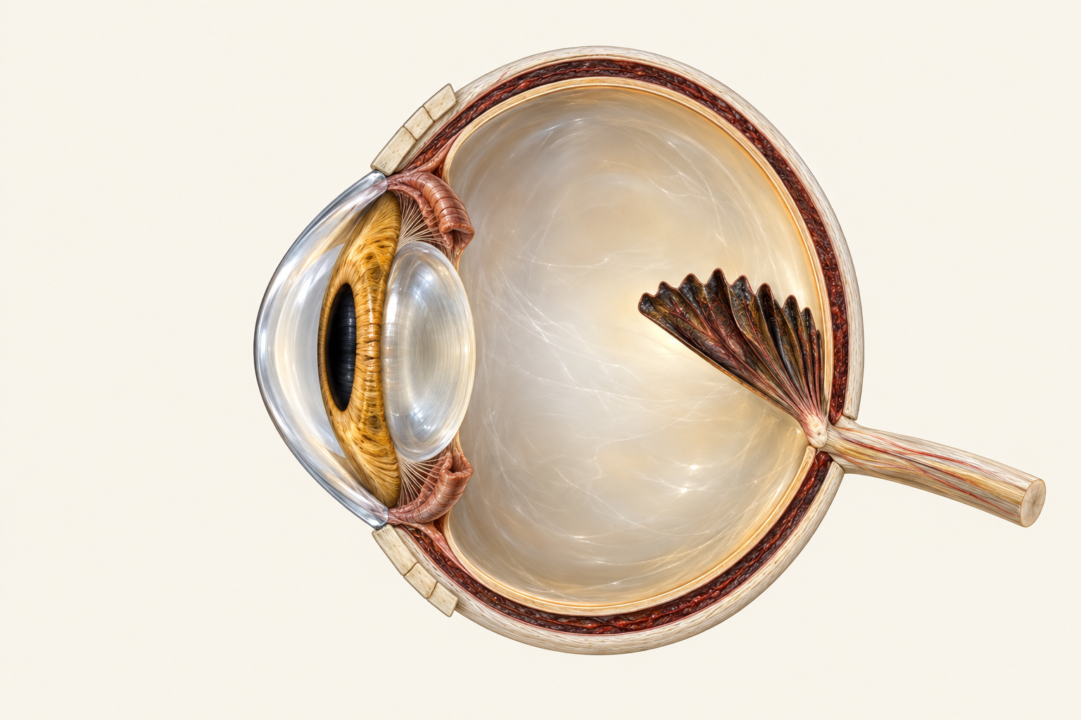
The bald eagle eye
Haliaeetus leucocephalus · ink & full colourSwitch between two detailed artistic studies. Enlarge the pecten, explore two foveae and their viewing directions, and move a translucent third eyelid.
explore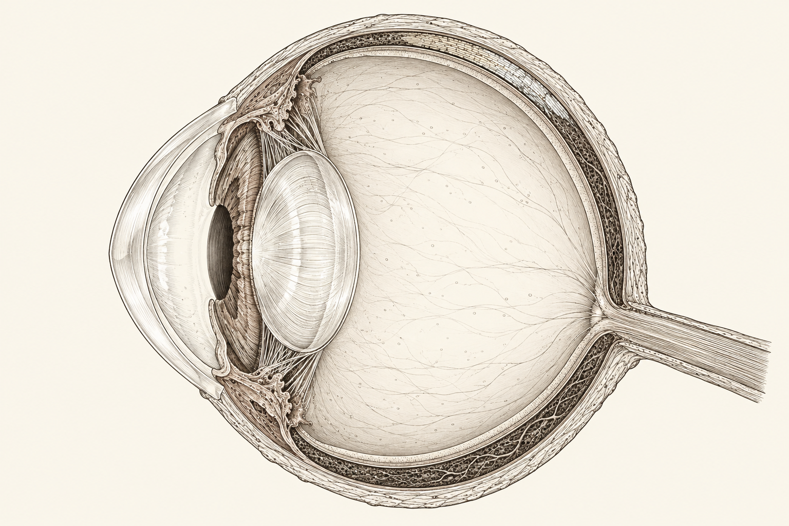
The domestic dog eye
Canis lupus familiaris — detailed inkInspect a fine ink cutaway, trace reflected light through a regional tapetum, find the area centralis and move a medial third eyelid.
explore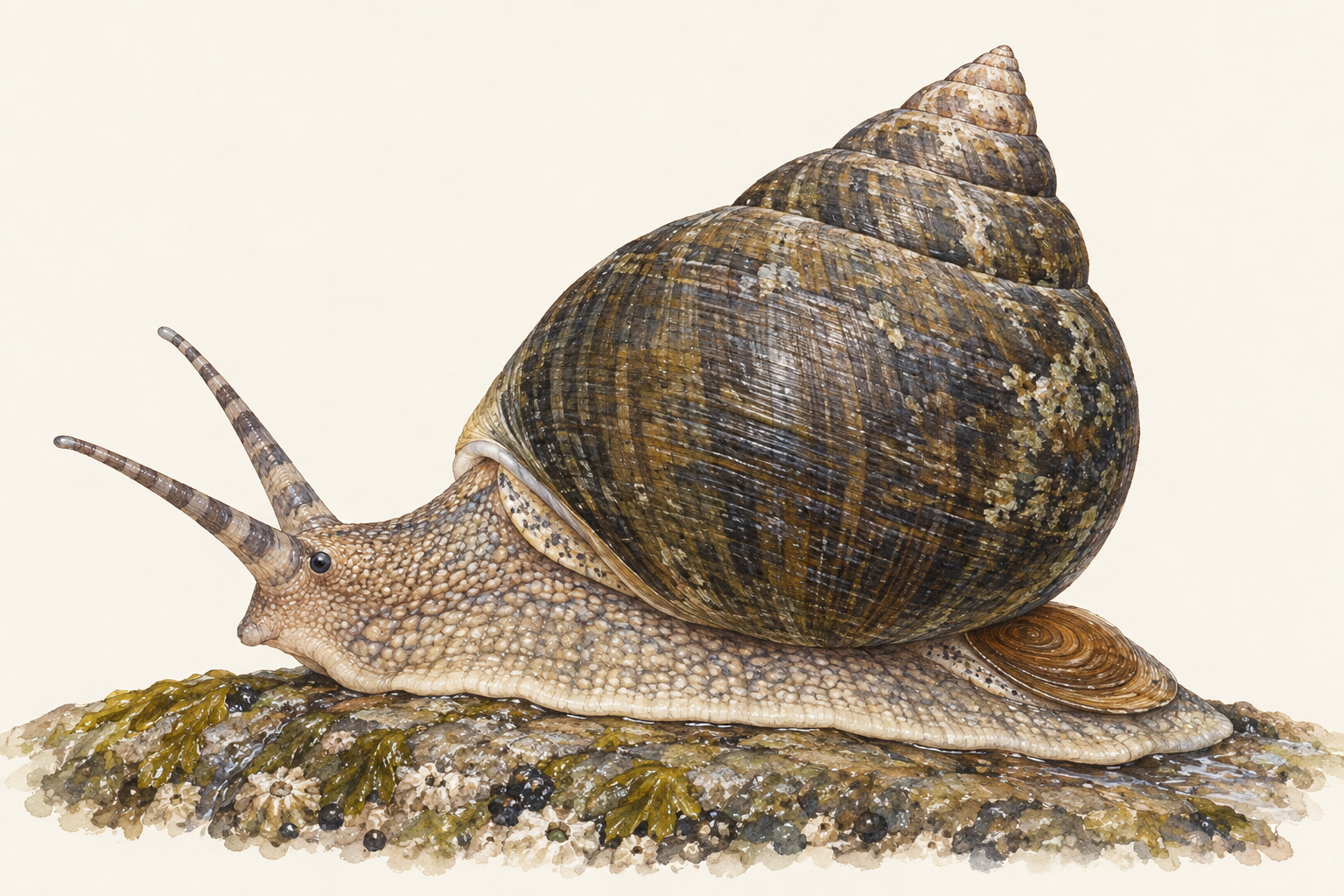
The common periwinkle
Littorina littorea — a natural-history studyFade a marine snail’s shell to inspect its body, compare a dextral aperture in two states, and magnify a grazer’s toothed ribbon.
explore →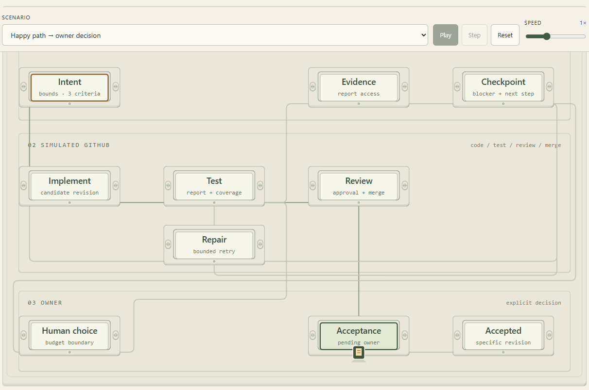
Can this change be called done?
A Workpacket-light policy experimentFollow a synthetic code change through tests, repair loops, stale evidence and checkpoints. A simulated merge still waits for explicit owner acceptance.
explore →the tools behind the field notes
These field notes are Adrian Hensler's experiments in interactive explanation, made with AI assistance and Anatomy, an open-source agent skill by Ryan (wheresryan22).
Anatomy supplies guidance and code for detailed isometric SVG figures, model-driven movement, optional WebGL effects and experimental 3D. Explore the source repository and original examples to see what the toolkit can do.
Our monarch study uses its page and frame helpers around a custom 2D drawing and label selector. It is a small first use of the toolkit, rather than a demonstration of its full capabilities. The butterfly illustration, text and implementation belong to this site's work; they are not an upstream Anatomy example. This independent adaptation implies no endorsement or scientific validation by the toolkit's creator. Biological sources are listed on the diagram page.
The upstream Anatomy code is released under the MIT License, copyright © 2026 Ryan. Our first study used this reviewed version.
Upstream MIT licence notice
MIT License

Copyright (c) 2026 Ryan

Permission is hereby granted, free of charge, to any person obtaining a copy
of this software and associated documentation files (the "Software"), to deal
in the Software without restriction, including without limitation the rights
to use, copy, modify, merge, publish, distribute, sublicense, and/or sell
copies of the Software, and to permit persons to whom the Software is
furnished to do so, subject to the following conditions:

The above copyright notice and this permission notice shall be included in all
copies or substantial portions of the Software.

THE SOFTWARE IS PROVIDED "AS IS", WITHOUT WARRANTY OF ANY KIND, EXPRESS OR
IMPLIED, INCLUDING BUT NOT LIMITED TO THE WARRANTIES OF MERCHANTABILITY,
FITNESS FOR A PARTICULAR PURPOSE AND NONINFRINGEMENT. IN NO EVENT SHALL THE
AUTHORS OR COPYRIGHT HOLDERS BE LIABLE FOR ANY CLAIM, DAMAGES OR OTHER
LIABILITY, WHETHER IN AN ACTION OF CONTRACT, TORT OR OTHERWISE, ARISING FROM,
OUT OF OR IN CONNECTION WITH THE SOFTWARE OR THE USE OR OTHER DEALINGS IN THE
SOFTWARE.